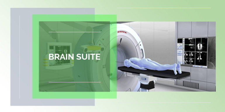
Brain Suite:
A Brain Suite is a cutting edge technology that allows our surgeons to effectively operate on brain tumors.
In this technique, the entire brain surgery will be performed under the guidance of intra operative high resolution MRI imaging. As the surgeon performs the brain surgery, live images will be displayed on the screen. This helps the surgeon fully remove the diseased part of the brain, without damaging the normal brain areas.
What is Brain Suite with Intra-operative Imaging?
Brain Suite with intra-operative MRI is a state of the art Neurosurgery operating theatre, which has the capability of intra-operative MR (magnetic resonance) imaging and MR-guided surgery. It helps surgeons to perform a high-resolution MRI during the surgery, vs. the regular scan which is done outside the operating room, which gives surgeons a real-time view of their progress during complex brain surgeries.
The image guidance during the surgery acts as a GPS (Global Positioning System) — it navigates the surgeon through the channels of the brain, so as to ensure the precision in the surgery. This prevents damage to normal brain tissues and also helps in minimizing the damage to the area from where tumor is resected. It helps in ensuring the nerves and important tracts are saved as much as possible.
What makes Brain Suite Intra-operative Imaging unique?
Brain Suite reduces the risk of damaging the crucial parts of the brain and help the surgery to be successful. It provides a complete check if some more resection is required before the closing of the patient’s head and completion of the surgery. Intraoperative Imaging is known to reduce the re-exploration (revised surgery) rates.
How does it help?
Image guidance helps in performing precise surgery and prevents damage to healthy brain tissues, thus helping in the preservation of normal brain functions. Intra-operative MRI helps in performing an immediate assessment of the degree of tumour removal. This ensures that adequate tumour removal is performed. MRI guidance helps in immediate detection of complications.
The difference between a normal surgery and Neuro-navigation in Brain Suite can be understood as driving with a GPS navigation system. Just as a GPS navigation system helps you in real-time through traffic routes and makes it easy for you to drive, similarly the Neuro-navigation attached to the Brain Suite helps your surgeon to get a real-time image of the tumor, as compared to a normal surgery where MRI is done outside the OT and then tumor is visible only after the skull is opened.
Preparing for Intra-operative imaging:
- The doctor would want to know if the patient has any metal implants or has worked with the sheet metal. If the patient is wearing anything related to metal or any jewellery, the doctor will ask him/her to remove it.
- The medical staff will ask the patient to wear a loose gown before the surgery. It is advisable to let the doctor know if the patient is pregnant.
During the Intra-operative imaging:
- Portable imaging devices, such as brain suites, are moved into the operating room to create images which are kept nearby, while the surgery is to be performed.
- The duration of the surgery depends on the requirement of the imaging of the patient’s brain and the condition of the patient’s tumours.
What are the benefits and risks of Brain Suite intra-operational imaging?
Intra-operational imaging is a non-invasive and painless test that results in high-resolution images of the brain within a short span of time.
Benefits:
- Greater precision is provided to the surgeon by integrating the MRI imaging and the operating room navigation to ensure that the complete tumour is removed without damaging any healthy tissues.
- Real time imaging is possible so the patient does not need to undergo multiple operations to check if the entire tumour has been removed.

